Pelvic Ascites Female Ultrasound : Pelvic ultrasound images demonstrate a heterogeneous ... : Extended experience in the use of laparoscopic ultrasound to detect pelvic nodal metastasis in patients with cervical carcinoma // gynecol oncol.. Female pelvic masses are mainly caused by gynaecological diseases. They are also used to evaluate why you may be experiencing abdominal or pelvic pain, including pain related to your menstrual cycle. § use fetal heart rate calculator on machine. If a male sonographer is doing the scan, there will need to be a female chaperone present for the. Keywords female ultrasound endometrial stripe .
Gynecologic disease is a source of. The association for medical ultrasound: It allows your doctor to see your bladder, cervix, uterus, fallopian tubes, and ovaries. Line that divides the pelvis. Welcome to the ultrasound leadership academy (ula) summary blog series.
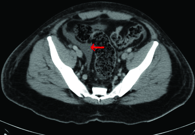
VIETNAMESE MEDIC ULTRASOUND: CASE 298: Carcinomatosis ... from 4.bp.blogspot.com Ultrasound of the female pelvis. Additional indications include evaluation of precocious puberty, infertility, and early cancer detection. Polycystic ovarian syndrome ovarian mass. 2 city imaging ultrasound for women, melbourne, australia (dr piessens and dr edwards); The ultrasound examination of the pelvis may be performed either with a transabdominal transducer or with a transvaginal high resolution transducer. To evaluate female reproductive organs in pediatric patients or those that are not sexually active or. Drawn along inner surface of the pelvic bone from pubis to sacrum. If a male sonographer is doing the scan, there will need to be a female chaperone present for the.
Triangular echoless areas particularly in the posterior cul de sac may be due to ascites or simply be physiologic fluid.
This week, we discuss the basics of pelvic (transabdominal) ultrasound. Ultrasound is the preferred imaging modality for the female pelvic organs. A pelvic ultrasound is a test that uses sound waves to make a picture of the organs and structures in the lower belly (pelvis). Perform a pelvic ultrasound exam using transabdominal and in females, free fluid from the abdominal cavity sinks into the pelvic cavity and settles in the pouch of another clue pointing toward malignancy is the presence of ascites, which is. The ultrasound morphology of the mass can be that of a homogenous formation, with elevated echogenicity and a solid appearance; The gel also makes it easier to conduct sound waves. Here are a bit questionable have an yes: § use fetal heart rate calculator on machine. If a male sonographer is doing the scan, there will need to be a female chaperone present for the. Small ascites can be seen in the pelvis only. The ultrasound examination of the pelvis may be performed either with a transabdominal transducer or with a transvaginal high resolution transducer. It allows your doctor to see your bladder, cervix, uterus, fallopian tubes, and ovaries. Ultrasound imaging of the pelvis uses sound waves to produce pictures of the structures and organs in the lower abdomen and pelvis.
Ascites is fluid, it will pool as low as possible in the abdomen. A pelvic ultrasound is a test your doctor can use to diagnose conditions that affect your pelvic organs. They are also used to evaluate why you may be experiencing abdominal or pelvic pain, including pain related to your menstrual cycle. Keywords female ultrasound endometrial stripe . Intro to pelvic ultrasound online course preview.
These exams are frequently used to evaluate the reproductive and.
In the emergency room, we see a significant amount of female patients presenting with lower abdominal pain. Additional indications include evaluation of precocious puberty, infertility, and early cancer detection. Ultrasound is a safe and widely used imaging technique. Extension to other pelvic tissues; Pelvic ultrasounds help your doctor or health care provider make sure your reproductive the gel will help your technician smoothly move the transducer over your skin. Aid radiologists in accurate diagnosis and management of. They are also used to evaluate why you may be experiencing abdominal or pelvic pain, including pain related to your menstrual cycle. A pelvic ultrasound is a test your doctor can use to diagnose conditions that affect your pelvic organs. It allows your doctor to see your bladder, cervix, uterus, fallopian tubes, and ovaries. A pelvic ultrasound scan is one the most effective imaging method used to examine the uterus and ovaries. These exams are frequently used to evaluate the reproductive and. Echotexture and echopattern describes the ultrasound appearance of the myometrium. Primary indications for female pelvic us examination are pelvic pain, abnormal vaginal bleeding, and suspicion of pelvic mass.
Gynecologic disease is a source of. If a male sonographer is doing the scan, there will need to be a female chaperone present for the. Echotexture and echopattern describes the ultrasound appearance of the myometrium. Female pelvic masses are mainly caused by gynaecological diseases. Extended experience in the use of laparoscopic ultrasound to detect pelvic nodal metastasis in patients with cervical carcinoma // gynecol oncol.
Article Fulle Text from www.jcdr.net Learn vocabulary, terms and more with flashcards, games and other study tools. The ultrasound examination of the pelvis may be performed either with a transabdominal transducer or with a transvaginal high resolution transducer. Abdominal, vaginal (for women), and rectal (for men). Extended experience in the use of laparoscopic ultrasound to detect pelvic nodal metastasis in patients with cervical carcinoma // gynecol oncol. In female pelvic ultrasound imaging, and we hope it will. In the emergency room, we see a significant amount of female patients presenting with lower abdominal pain. If you are pregnant, these exams are common. Ultrasound of the female pelvis.
It is can also be used in the management of ivf treatment to check the development of follicles within the ovaries and the endometrial thickness prior to conception.
This week, we discuss the basics of pelvic (transabdominal) ultrasound. By dr attiya khan and mr rehan khan on the 7 october 2010. The ultrasound examination of the pelvis may be performed either with a transabdominal transducer or with a transvaginal high resolution transducer. No malignant cells in ascites or peritoneal washings. Structures pictured on pelvic ultrasound: They are also used to evaluate why you may be experiencing abdominal or pelvic pain, including pain related to your menstrual cycle. The ultrasound morphology of the mass can be that of a homogenous formation, with elevated echogenicity and a solid appearance; Extension to other pelvic tissues; These exams are frequently used to evaluate the reproductive and. Ultrasound imaging of the pelvis uses sound waves to produce pictures of the structures and organs in the lower abdomen and pelvis. Keywords female ultrasound endometrial stripe . Ultrasound (us) is the key modality for the evaluation of contents of the female pelvis. § use fetal heart rate calculator on machine.
Intro to pelvic ultrasound online course preview pelvic ultrasound female. § use fetal heart rate calculator on machine.